AIR Scientists Develop Hyperspectral Surface Plasmon Resonance Microscopy
Hyperspectral surface plasmon resonance microscopy (HSPRM) is an advanced analytical technique for spectral imaging and chemical and biological sensing, which enables high-resolution visualization and precise quantification of chemical and biological analytes.
A study published in Nature Communications describes a flexible HSPRM system that operates by using a hyperspectral microscope to analyze the selected area of SPR image produced by a prism-based spectral SPR sensor.
The HSPRM system is developed by a research team of the State Key Laboratory of Transducer Technology at the Aerospace Information Research Institute (AIR), Chinese Academy of Sciences (CAS).
The HSPRM system enables monochromatic and polychromatic SPR imaging and single-pixel spectral SPR sensing, as well as two-dimensional quantification of thin films with the measured resonance-wavelength images. It can measure SPR radiance spectra instead of conventional intensity spectra to improve the figure of merit (FOM) of single-pixel spectral SPR sensors and can also quantify two-dimensional profiles of thickness and refractive index for thin films by using the measured resonance-wavelength images.
Pixel-by-pixel calibration of the incident beam collimation deviation was performed to remove pixel-to-pixel differences in SPR sensitivity.
The HSPRM system has a wide spectral range from 400 nm to 1000 nm, an optional field of view from 0.884 mm2 to 0.003 mm2 and a high lateral resolution of 1.2μm.
Several typical applications of the HSPRM system are shown in this study, including quantification of single-layer graphene thickness distribution, in situ detection of inhomogeneous protein adsorption, and label-free single cell analysis.
The system, marking an innovative breakthrough in SPR sensor technology, is expected to greatly expand the SPR imaging applications.
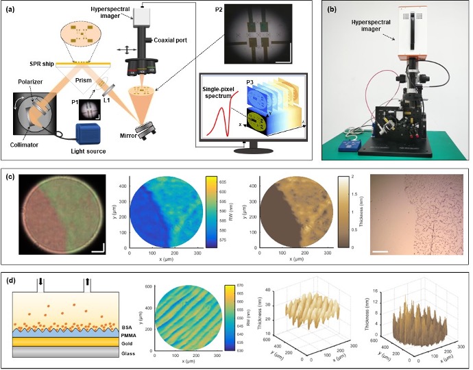
Figure 1 (a) Schematic diagram of the HSPRM system. (b) Photograph of the HSPRM system constructed in our laboratory. (c) Quantification of monolayer graphene thickness using the HSPRM system. From left to right: spectral SPR image of the monolayer graphene covered SPR chip, RW image extracted from the measured hyperspectral datacube, thickness map of the monolayer graphene, optical microscope image of the same graphene sample. (d) Detection of inhomogeneous protein adsorption. From left to right: schematic diagram of BSA adsorption on a wrinkled PMMA film, RW image of the wrinkled PMMA film, 2D thickness distribution of the wrinkled PMMA film, 2D distribution of the BSA adlayer thickness.(Image by AIR)
News & Events